Back تهجين موضعي متألق Arabic Fluorescentna ''in situ'' hibridizacija BS Hibridació in situ per fluorescència Catalan Fluorescenční in situ hybridizace Czech FISH-Test German Μέθοδος FISH Greek Hibridación fluorescente in situ Spanish Hybridation in situ en fluorescence French Hibridación in situ fluorescente Galician היברידיזציה פלואורסצנטית באתר HE


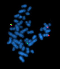
Fluorescence in situ hybridization (FISH) is a molecular cytogenetic technique that uses fluorescent probes that bind to only particular parts of a nucleic acid sequence with a high degree of sequence complementarity. It was developed by biomedical researchers in the early 1980s[1] to detect and localize the presence or absence of specific DNA sequences on chromosomes. Fluorescence microscopy can be used to find out where the fluorescent probe is bound to the chromosomes. FISH is often used for finding specific features in DNA for use in genetic counseling, medicine, and species identification.[2] FISH can also be used to detect and localize specific RNA targets (mRNA, lncRNA and miRNA)[citation needed] in cells, circulating tumor cells, and tissue samples. In this context, it can help define the spatial-temporal patterns of gene expression within cells and tissues.
- ^ Langer-Safer PR, Levine M, Ward DC (July 1982). "Immunological method for mapping genes on drosophila polytene chromosomes". Proceedings of the National Academy of Sciences of the United States of America. 79 (14): 4381–4385. Bibcode:1982PNAS...79.4381L. doi:10.1073/pnas.79.14.4381. PMC 346675. PMID 6812046.
- ^ Amann R, Fuchs BM (May 2008). "Single-cell identification in microbial communities by improved fluorescence in situ hybridization techniques". Nature Reviews. Microbiology. 6 (5): 339–348. doi:10.1038/nrmicro1888. PMID 18414500. S2CID 22498325.